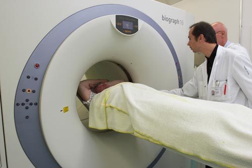
Right ventricular dilatation (RVD), as assessed by multidetector computed tomography (MDCT), is not associated with worse prognosis in patients with low risk pulmonary embolism (PE), according to a study published in the ERJ.
Researchers sought to investigate whether RVD assessed by MDCT in low-risk PE patients is associated with an increased risk of adverse outcomes in patients with a simplified Pulmonary Embolism Severity Index (sPESI) score of zero.
Benoit Côté and colleagues pooled analysis of 779 PE patients from three prospective cohorts from the PREP study, the PROTECT study and a French prospective single-centre registry.
The requirement to have a sPESI score of zero ruled outpatients with a history of cancer or cardiopulmonary disease, as well as those aged over 80 years, those with low oxygen saturation or systolic blood pressure, or elevated heart rate on admission.
The researchers found that the risk of PE-related adverse events and all-cause mortality during the first 30 days was low; PE-related adverse events occurred in 0.77% of patients, while all-cause mortality occurred in just 0.39% of the cohort [all deaths being attributed to PE].
The researchers conclude that the study confirms that RVD assessed by MDCT is not associated with worse prognosis, but cite several limitations including the method of retrospective analysis and the low number of events they were able to analyse.
The article is accompanied by an editorial, also published in the ERJ, by Dr. Mareike Lankeit, entitled ‘Always think of the right ventricle, even in “low-risk” pulmonary embolism’.


